√100以上 e coli gram stain morphology 230626-E coli gram stain morphology
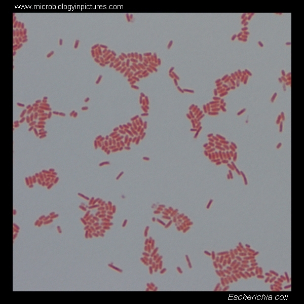
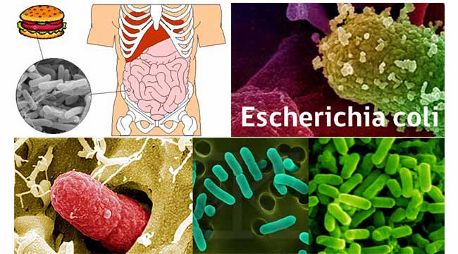
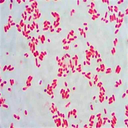
Escherichia coli is first isolated by Escherich in 15 Escherichia coli is the most important species encountered as a human pathogen Most commonly found in human and animal intestine There is various form of Escherichia coli present in nature Morphology of Escherichia coli Escherichia coli is a gramnegative, nonsporing, bacillusEscherichia coli is a nonsporeforming, Gramnegative bacterium, usually motile by peritrichous flagellaEscherichia coli is the most common cause of acute urinary tract infections as well as urinary tract sepsis It has also been known to cause neonatal meningitis and sepsis and also abscesses in a number of organ systems8 Mechanisms of enterotoxins 4 Escherichia coli (EHEC) 1 Gram Stain 2 Morphology 3 Special characteristics/virulence factors 4 Transmission 5 Exotoxins 6 MacConkey agar

Gram Stain Images Microbiology Stain Study Tools
E coli gram stain morphology
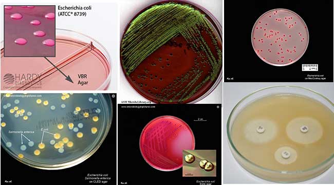
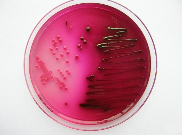
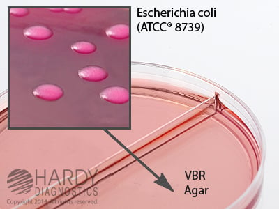
E coli gram stain morphology-Escherichia coli Four different strains of Escherichia coli on Endo agar with biochemical slope Glucose fermentation with gas production, urea and H 2 S negative, lactose positive (with exception of strain D "late lactose fermenter";E coli are Gramnegative bacteria, meaning that they do not retain the crystal violet stain commonly used to differentiate bacteria Their status as Gramnegative bacteria is due to their thin cell walls E coli has cell walls made out of two thing peptidoglycan layers, an inner and outer membrane


Q Tbn And9gcqkye60ou Johpr02n Mbv1fferrjpdh Lnct7ymdf5qhyia1ld Usqp Cau
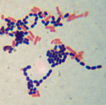
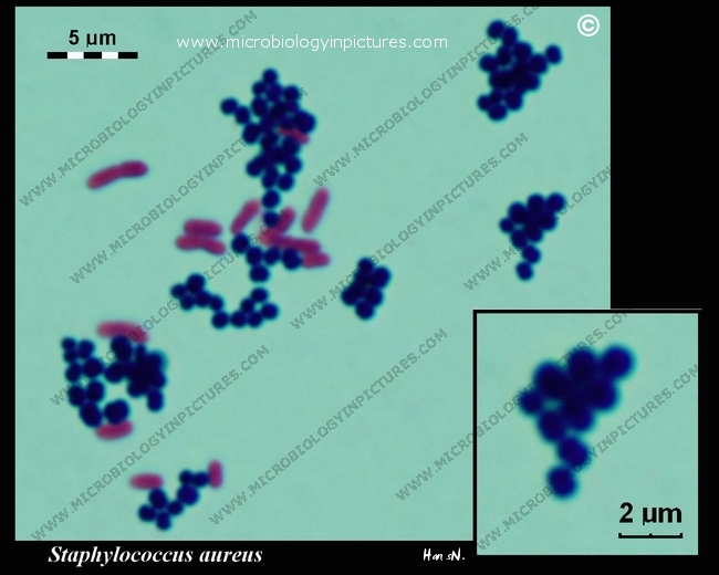
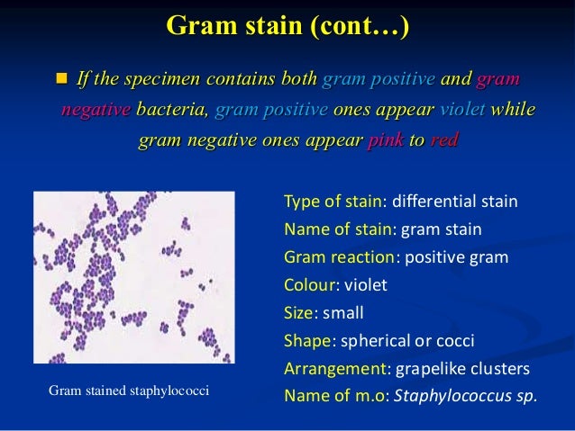
Your unknown sample For example, when you perform a Gram stain, you will always include samples of Staphylococcus epidermidis (S epi), which is known to be Gram positive, and Escherichia coli, which is known to be Gramnegative If the Gram stain procedure works as it should, S epi will be purple and E coli will be pinkThrough this experiment, gram staining skills develop More understanding the types and morphology of bacteria Expected experimental result, Escherichia coli (Ecoli ) is a negative gram bacteria which stain pink colour , while Staphylococcus aureus ( Saureus ) is a positive gram bacteria which stain purple colour Materials BacteriaIn Atlas of Oral Microbiology, 15 315 Staphylococcus Members of the Staphylococcus genus are grampositive cocci and belong to the Micrococcus family The organisms are widely spread in the environment Early on, three species were isolated from clinical samples Staphylococcus aureus, S epidermidis, and S saprophyticusIn the early 1980s, analysis of biochemical reactions (eg
Escherichia coli Four different strains of Escherichia coli on Endo agar with biochemical slope Glucose fermentation with gas production, urea and H 2 S negative, lactose positive (with exception of strain D "late lactose fermenter";THE PROCEDURE done individually We have cultures of E coli and Bacillus for you to gram stainThis will give you gram and gram – controls to check your procedure against You can use 2 slides, 1 for each bacterium, or you can divide one slide in half and smear each bacterium on the divided slideTheir DNA forms a tangle known as a nucleoid, but there is no membrane around the
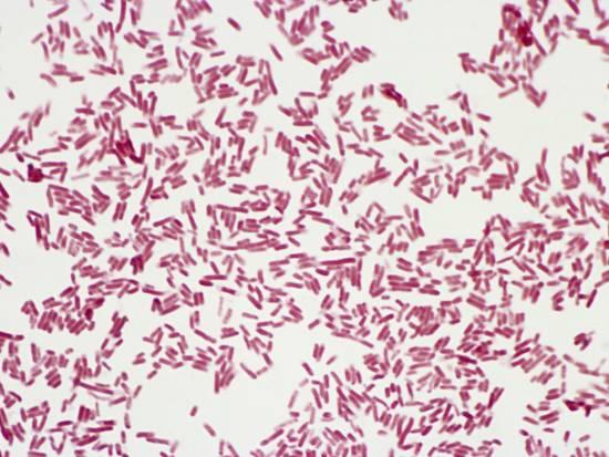
The Gram stain is a differential staining technique used to classify & categorize bacteria into two major groups Gram positive and Gram negative, based on the differences of the chemical and physical properties of the cell wall Escherichia coli Fig Gram negative bacteria Bacterial Morphology Bacteria are very small unicellularOn staining, E coli appear as nonsporeforming, Gramnegative rodshaped bacterium;Their DNA forms a tangle known as a nucleoid, but there is no membrane around the


Salmonella And Salmonellosis
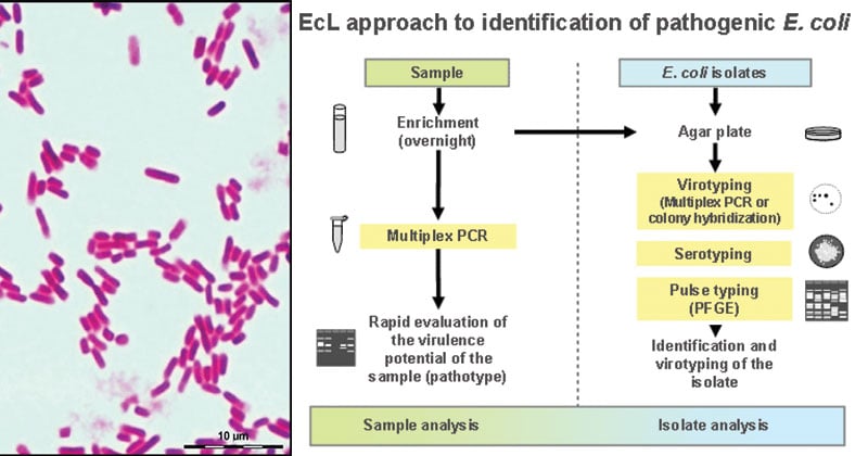


Escherichia Coli E Coli An Overview Microbe Notes
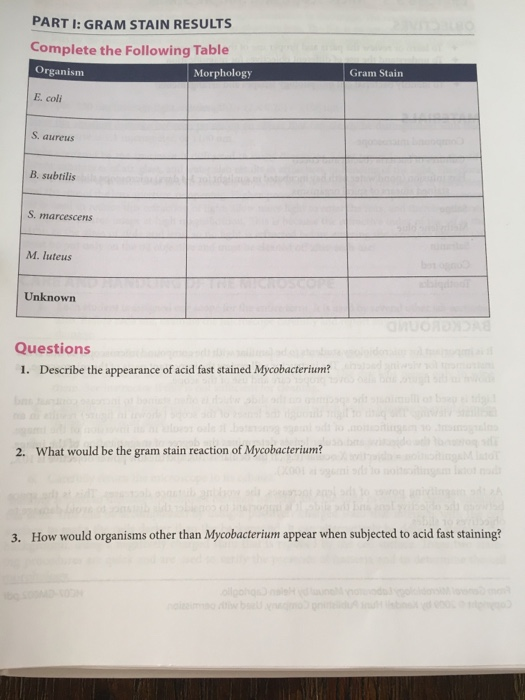
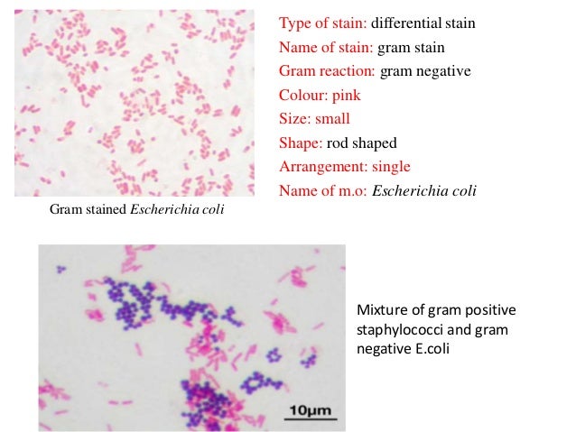
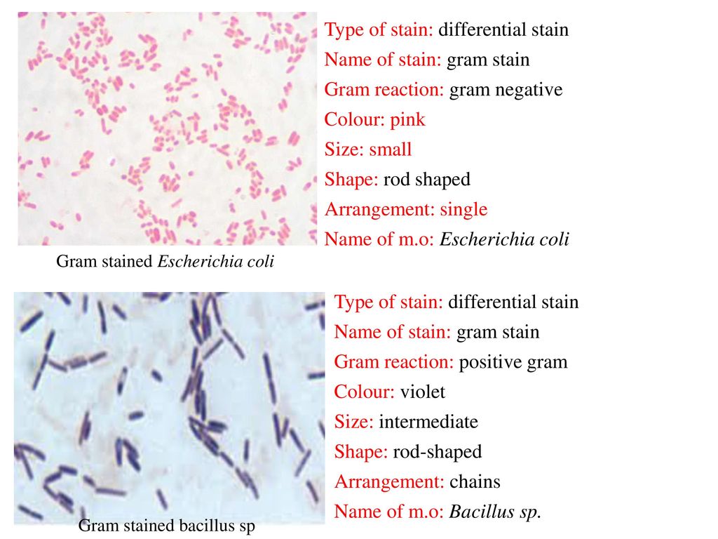
Ex 36 Gram Stain OBSERVATIONS AND INTERPRETATIONS Record your observations in the chart below Cellular Morphology and Arrangement (Include a detailed sketch of a few representative cells) Gram Reaction (/) Organism or Source Color Mention the shape and arrangement of E coli Mention E coli color Mention Gram nature of E coli E coli and S epidermidis mixed Mention the shape andCells are typically rodshaped, and are about μm long and 025–10 μm in diameter, with a cell volume of 06–07 μm 3 E coli stains Gramnegative because its cell wall is composed of a thin peptidoglycan layer and an outer membraneE coli S epidermidis Note Escherichia coli is a tiny pink (Gram) rod Staphylococcus epidermidis is a purple (Gram) sphere or coccus Draw a picture of a typical microscopic field and identify both Escherichia coli and Staphylococcus epidermidis Record this in the results section for this lab Colored pencils are available throughout the room
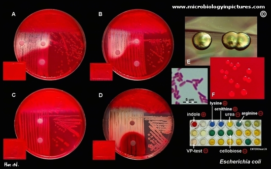


Escherichia Coli Colony Morphology And Microscopic Appearance Basic Characteristic And Tests For Identification Of E Coli Bacteria Images Of Escherichia Coli Antibiotic Treatment Of E Coli Infections


Gram Negative Bacteria Wikipedia
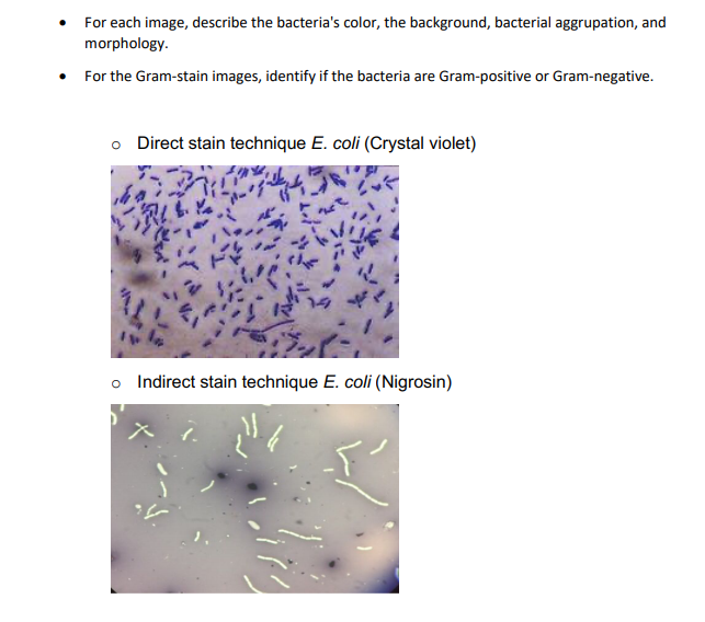
It was devised originally by a Danish bacteriologist, Hans Christian Joachim Gram (14) as a method of staining bacteria in his laboratory Morphology of these bacteria are Gram negative rods measuring 13 µm × 0407 µm Most strains are motile by peritrichates flagella They are nonsporing and non encapsulated Principle of Gram stainGramstain Negative = 0, Positive = 1, Indeterminate = 2 Found in human microbiome Microbes that live anywhere in the human body and are not pathogenic to humans (ie capable of causing human disease) No = 0, Yes = 1 Plant pathogen Does the species causes disease in plants?On the other hand, the differential stains are Gram stain and endospore stain Escherichia coli was used to stain with a basic dye, crystal violet and the result was shown in Figure 1 As shown in the figure, the cells of Escherichia coli were purple colour and rodshaped as their appearances


Lab 1


Sem Images Of Gram Negative Bacteria Including E Coli And P Download Scientific Diagram
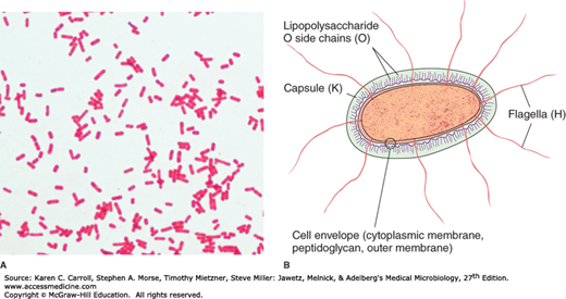
Common examples of gramnegative include Salmonella spp, Escherichia coli (E coli) and the Enterobacteriaceae spp GramPositive Bacteria Unlike gramnegative, gram positive bacteria have a thick peptidoglycan layer that allows them to retain the primary stain/dye (crystal violet stain)MORPHOLOGY OF ESCHERICHIA COLI (E COLI) Shape – Escherichia coli is a straight, rod shape (bacillus) bacterium Size – The size of Escherichia coli is about 1–3 µm × 04–07 µm (micrometer) Arrangement Of Cells – Escherichia coli is arranged singly or in pairs Motility – Escherichia coli is a motile bacteriumType and morphology E coli is a Gramnegative, facultative anaerobe (that makes ATP by aerobic respiration if oxygen is present, but is capable of switching to fermentation or anaerobic respiration if oxygen is absent) and nonsporulating bacterium Cells are typically rodshaped, and are about μm long and 025–10 μm in diameter, with a cell volume of 06–07 μm 3
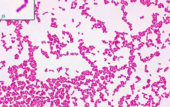


Escherichia Coli O157 H7


Www Mccc Edu Hilkerd Documents Bio1lab3 Exp 4 Pdf
E coli is described as a Gramnegative bacterium This is because they stain negative using the Gram stain The Gram stain is a differential technique that is commonly used for the purposes of classifying bacteriaPercentage of isolation of the five E coli Colonial Morphology (CM) classes from inpatients and outpatients (EMB) agar, also the result of Gram staining agrees with the findings of the studyEscherichia coli (EPEC) 1 Gram Stain 2 Morphology 3 Special characteristics/virulence factors 4 Transmission 5 Exotoxins 6 MacConkey agar results 7 Invasive?
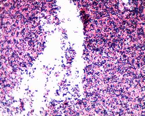


Gram Stain Microbiology Images Photographs


Staphylococcus Aureus And Ecoli Under Microscope Microscopy Of Gram Positive Cocci And Gram Negative Bacilli Morphology And Microscopic Appearance Of Staphylococcus Aureus And E Coli S Aureus Gram Stain And Colony Morphology On Agar Clinical
Gram stain or Gram staining, also called Gram's method, is a method of staining used to distinguish and classify bacterial species into two large groups grampositive bacteria and gramnegative bacteriaThe name comes from the Danish bacteriologist Hans Christian Gram, who developed the technique Gram staining differentiates bacteria by the chemical and physical properties of their cell wallsEscherichia coli, often abbreviated E coli, are rodshaped bacteria that tend to occur individually and in large clumps E coli bacteria have a single cell arrangement, according to Schenectady County Community College E coli is a gramnegativRoutine urine cultures should be plated using calibrated loops for the semiquantitative method Note The most commonly used criterion for defining significant bacteriuria is the presence of ⩾10 5 CFU per milliliter of urine


Welcome To Microbugz Gram Stain


Enteric Gram Negative Rods Enterobacteriaceae Basicmedical Key
Escherichia coli or in short E coli is a rodshaped microorganism It is categorized as gramnegative based on the gram staining procedure E coli is usually found in the large intestine of mammals and is the most abundant microorganism to be present in the feces Hence, it is used as an indicator for water purityGram Staining Now that the slide has been heat fixed, you may now begin to stain the organism This is started by applying a few drops of crystal violet for 30 seconds, then rinsing with water for 5 seconds Next, cover the slide with Gram's Iodine and let sit for a full minute before rinsing with water for another 5 secondsGram stain morphology for EscherichiaEscherichia coli Gramnegative Short rods (bacilli) Encapsulated and Unencapsulated Gram stain morphology for FrancisellaFrancisella tularensis


Performance Validation Of Chromogenic Coliform Agar For The Enumeration Of Escherichia Coli And Coliform Bacteria Lange 13 Letters In Applied Microbiology Wiley Online Library


Escherichia Coli Wikipedia
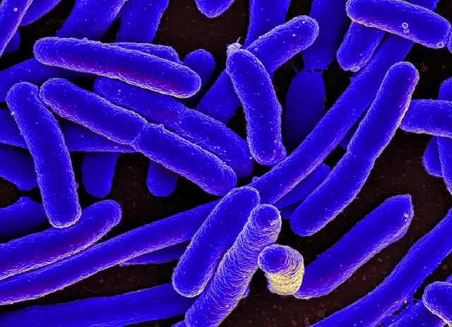
Staphylococci (staphylococcus aureus)gram positive, spherical bacteris that have developed antibiotic resistant strains, 400x gram stain stock pictures, royaltyfree photos & images microscopic view of bacterial pneumonia bacterial pneumonia is a type of pneumonia caused by bacterial infection gram stain stock illustrationsWe have cultures of E coli and Bacillus for you to gram stain This will give you gram and gram – controls to check your procedure against You can use 2 slides, 1 for each bacterium, or you can divide one slide in half and smear each bacterium on the divided slide There are also prepared, gram stained slides of bacteria of different shapes and sizes of bacteria to look atBacteria More on Morphology A more or less typical bacterium, shown here, is comparatively much simpler than a typical eukaryotic cell View the transmission electron micrograph of a typical bacterium, E coli, below and compare it with the diagram above Bacteria lack the membranebound nuclei of eukaryotes;


E Coli Under The Microscope Types Techniques Gram Stain Hanging Drop Method


Gram Positive Bacteria Microbiology
Grampositive cells, like Staphylococcus aureus, are purple under the microscope and Gramnegative cells, for example Escherichia coli or Neisseria subflava, turn out red The acid fast stain differentiates bacterial cells with lipoidal cell callGram stain morphology for EscherichiaEscherichia coli Gramnegative Short rods (bacilli) Encapsulated and Unencapsulated Gram stain morphology for FrancisellaFrancisella tularensisOn Endo agar it looks like lactose negative)All four strains are mannitol positive (best seen in fig D), cellobiose negative (strains A, B)


Colony Characteristics Of E Coli Sciencing


Escherichia Coli E Coli An Overview Microbe Notes
The colony morphology of organisms is observed Colony shape, size (in mm), color, consistency, elevation, opacity, and margin are observed and noted down for further identification Gram staining for identifying gram negative bacteria Gram staining is the beginning test in an identification procedure in bacterial classificationTHE PROCEDURE done individually We have cultures of E coli and Bacillus for you to gram stainThis will give you gram and gram controls to check your procedure against You can use 2 slides, 1 for each bacterium, or you can divide one slide in half and smear each bacterium on the divided slideGram Staining reaction – Gram ve uniform turbidity is produced which is further analyzed for the morphology (under the microscope), gram reaction, biochemical tests, and staphylococcus specific tests That's all about the Morphology & Cultural Characteristics of Staphylococcus aureus


Solved 2 10mm Figure 1 Gram Stain Bacteria Are Escherich Chegg Com


Gram Negative Rods Ppt Download
Positive Gram Stain Negative E coli Serratia marcescens Proteus mirabilis Neisseria gonorrhea Alcaligenes Faecalis Proteus vulgaris Klebsiella pneumoniae Citrobacter freundii Pseudomonas fluorescens Enterobacter aerogenes Staphylococcus aureus Streptococcus pneumoniae Bacillus cereus Enterococcus faecalis Micrococcus luteus E coli SerratiaOn Endo agar it looks like lactose negative)All four strains are mannitol positive (best seen in fig D), cellobiose negative (strains A, B)Bacteria More on Morphology A more or less typical bacterium, shown here, is comparatively much simpler than a typical eukaryotic cell View the transmission electron micrograph of a typical bacterium, E coli, below and compare it with the diagram above Bacteria lack the membranebound nuclei of eukaryotes;


Www Wiv Isp Be Qml Activities External Quality Rapports Atlas Bacteriology Gram Negative Aerobic And Facultative Rods Pdf


E Coli Bacteria Britannica
Bacteria More on Morphology A more or less typical bacterium, shown here, is comparatively much simpler than a typical eukaryotic cell View the transmission electron micrograph of a typical bacterium, E coli, below and compare it with the diagram above Bacteria lack the membranebound nuclei of eukaryotes;Microorganisms are very small microscopic structures that are capable of free living Some of the microorganisms are nonpathogenic and live on the body of human beings ie on the skin, in the nostrils, in the intestinal tract etc, and they are called commensals The organisms that are capable of causing disease are called pathogenic organisms These are two groups depending upon theTheir DNA forms a tangle known as a nucleoid, but there is no membrane around the


Gram Stain Images Microbiology Stain Study Tools


Gram Stain
Based on Staining reaction • GRAM'S STAIN – Grampositive cocci – Staphylococcus aureus – Gramnegative cocci – Neisseria gonorrhoeae – Grampositive rods – Clostridium spp – Gramnegative rods – E coli • ACID FAST STAIN – Acidfast bacilli –Mycobacterium tuberculosis – Nonacidfast bacilli – StaphylococcusDifferential stains Gram stain In contrast to simple stains, differential stains are used to distinguish the difference between bacteria One of the most wellknown differential stains is Gram stain, which differentiates grampositive and gramnegative bacteria based on the difference in their cell wall structure The Gram stain was developed by the Danish bacteriologist Hans Christian GramEscherichia coli or in short E coli is a rodshaped microorganism It is categorized as gramnegative based on the gram staining procedure E coli is usually found in the large intestine of mammals and is the most abundant microorganism to be present in the feces Hence, it is used as an indicator for water purity


E Coli Gram Stain And Cell Morphology E Coli Micrograph Appearance Under The Microscope E Coli Cell Morphology E Coli Microscopic Picture


Q Tbn And9gcqkye60ou Johpr02n Mbv1fferrjpdh Lnct7ymdf5qhyia1ld Usqp Cau
The Gram stain is a differential staining technique used to classify & categorize bacteria into two major groups Gram positive and Gram negative, based on the differences of the chemical and physical properties of the cell wall Escherichia coli Fig Gram negative bacteria Bacterial Morphology Bacteria are very small unicellularNo = 0, Yes = 1 Animal pathogen Does the species causes diseaseGram positive organisms absorb the stain and appear blue or purple under the microscope The gram positive S Pneumoniae is the most common cause of the typical community acquired pneumonia Staph aureus organisms are the most common cause of nosocomial infections overall (wound and skin)


Pathogenic E Coli


Escherichia Coli E Coli An Overview Microbe Notes
Staining procedures and to compare morphological features, such as size & shape of various microbes Today 1 Wet Mount 2 Heat Fixation required prior to staining 3 Simple Stain 4 Gram Stain 5 Review Stains Endospore, Capsule & AcidFast StainsE coli S epidermidis Note Escherichia coli is a tiny pink (Gram) rod Staphylococcus epidermidis is a purple (Gram) sphere or coccus Draw a picture of a typical microscopic field and identify both Escherichia coli and Staphylococcus epidermidis Record this in the results section for this lab Colored pencils are available throughout the roomEscherichia coli is first isolated by Escherich in 15 Escherichia coli is the most important species encountered as a human pathogen Most commonly found in human and animal intestine There is various form of Escherichia coli present in nature Morphology of Escherichia coli Escherichia coli is a gramnegative, nonsporing, bacillus


Www Mccc Edu Hilkerd Documents Bio1lab3 Exp 4 Pdf


Bacterial Characteristics Gram Staining Video Khan Academy
2 Morphology and Staining of Escherichia Coli E coli is Gramnegative straight rod, 13 µ x 0407 µ, arranged singly or in pairs (Fig 281) It is motile by peritrichous flagellae, though some strains are nonmotile Spores are not formed Capsules and fimbriae are found in some strains 3 Cultural Characteristics of Escherichia Coli


Learn About Discovery Of Escherichia Coli Chegg Com


Www Mccc Edu Hilkerd Documents Bio1lab3 Exp 4 Pdf


E Coli Gram Stain Mikrobiologiya


Www Wiv Isp Be Qml Activities External Quality Rapports Atlas Bacteriology Gram Negative Aerobic And Facultative Rods Pdf


Escherichia Coli E Coli An Overview Microbe Notes


Optical Microscope Images Of E Coli Cells Following Gram Staining A Download Scientific Diagram


Bacteria Exercise Flashcards Quizlet


Escherichia Coli Bacteria Appearance Dangerous Infections And Diseases E Coli O157 H7 Treatment Organs And Body Parts Affected By E Coli O157 H7 Bacteria Cell Morphology And Gram Stain Gram Negative Bacteria


Staphylococcus Aureus And Ecoli Under Microscope Microscopy Of Gram Positive Cocci And Gram Negative Bacilli Morphology And Microscopic Appearance Of Staphylococcus Aureus And E Coli S Aureus Gram Stain And Colony Morphology On Agar Clinical


A Representative Images Showing The Appearance Of Morphological Download Scientific Diagram


Yersinia Gram Stain Sd Dept Of Health


Pbstatemicrobiology Licensed For Non Commercial Use Only Gram Stain


Solved Part I Gram Stain Results Complete The Following Chegg Com


Gram Stain Of E Coli Bacterium A Gram Stain Of Shows Gramnegative Download Scientific Diagram


What Is The Cell Morphology Of Escherichia Coli Quora


Escherichia Coli Wikipedia


Q Tbn And9gcqkye60ou Johpr02n Mbv1fferrjpdh Lnct7ymdf5qhyia1ld Usqp Cau
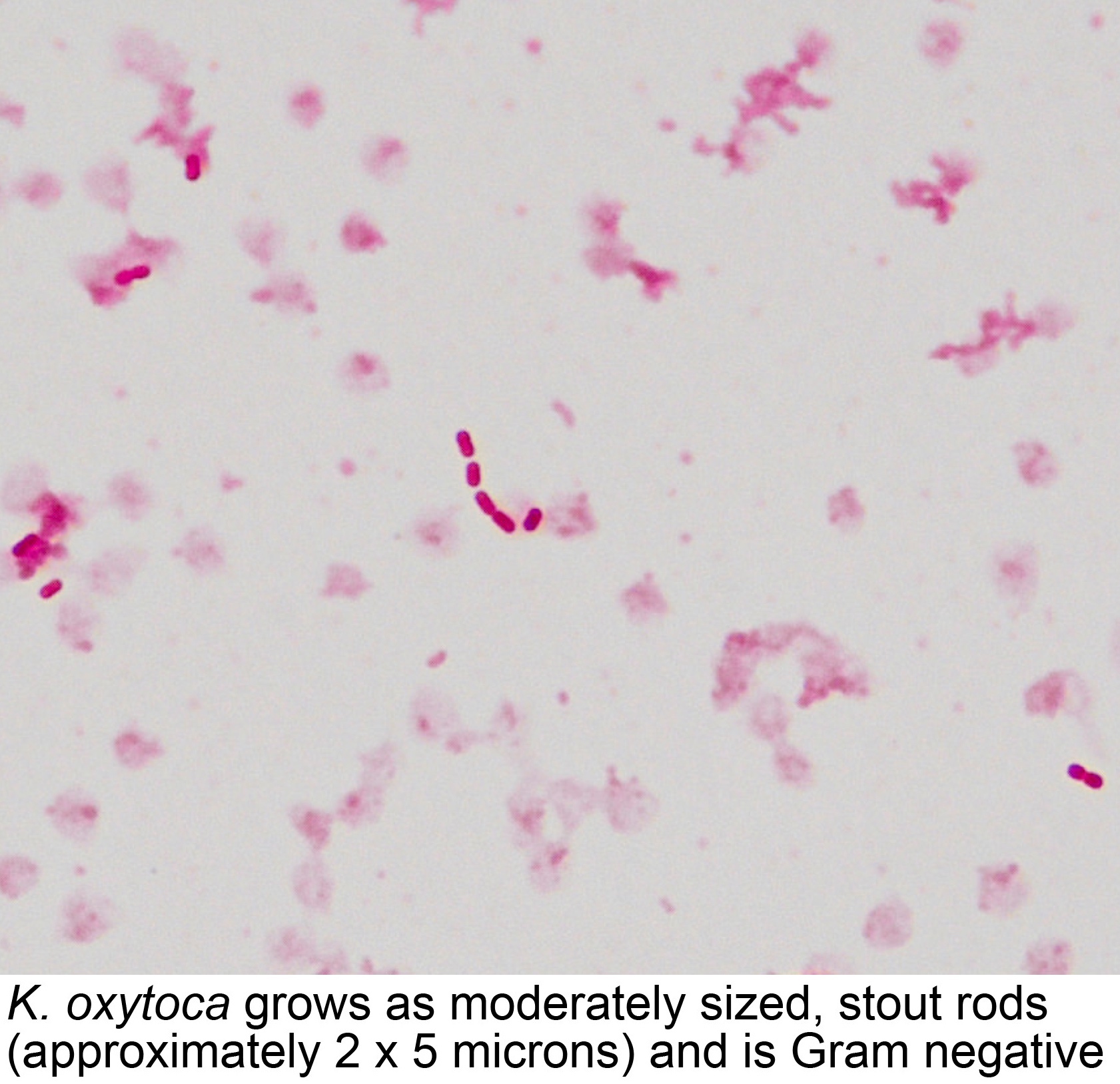


Pathology Outlines Klebsiella Oxytoca
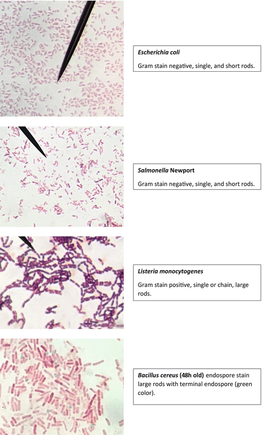


Staining Technology And Bright Field Microscope Use Springerlink
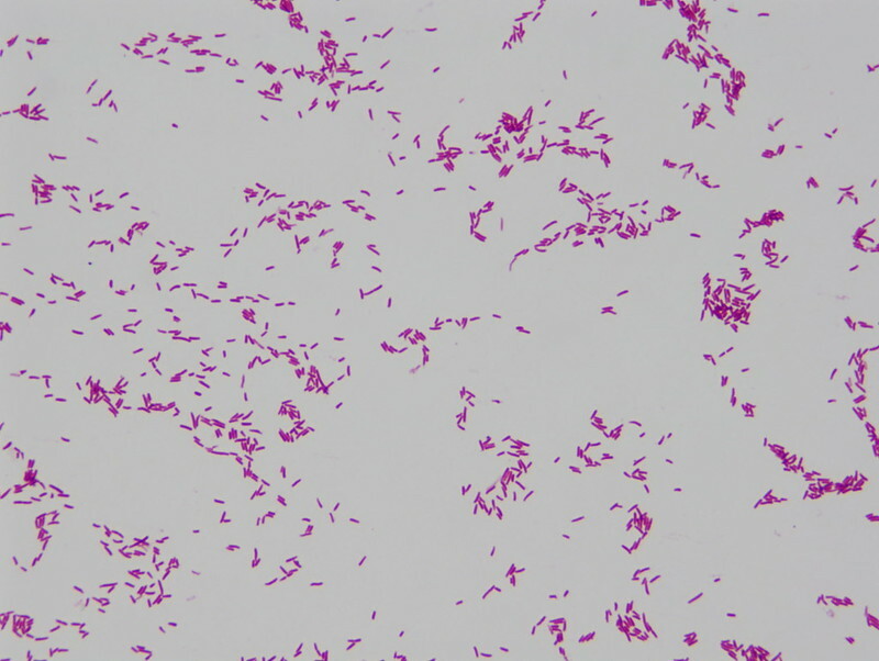


Pulsenotes Gram Negative Infections Notes


Academic Oup Com Labmed Article Pdf 32 7 368 Labmed32 0368 Pdf


Biol 230 Lecture Guide Gram Stain Of A Mixture Of Gram Positive And Gram Negative Bacteria


Optical Microscope Images Of E Coli Cells Following Gram Staining A Download Scientific Diagram


Gram Stain Wikipedia


What Does An E Coli Bacteria Look Like Under A Microscope Quora


Gram Stain Of Enterobacteria A Klebsiella G Emb B Salmonella Download Scientific Diagram


Gram Stain I Did To Check Morphology For My Labs B Meg And E Coli Microporn


Www Mccc Edu Hilkerd Documents Bio1lab3 Exp 4 Pdf
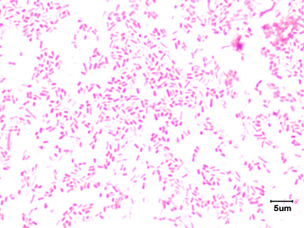


2 3b The Gram Negative Cell Wall Biology Libretexts


Bacterial Staining


Escherichia Coli Bacteria Appearance Dangerous Infections And Diseases E Coli O157 H7 Treatment Organs And Body Parts Affected By E Coli O157 H7 Bacteria Cell Morphology And Gram Stain Gram Negative Bacteria


Solved 1 Identify The Morphology Morphological Arrangem Chegg Com


Escherichia Coli Bacteria Appearance Dangerous Infections And Diseases E Coli O157 H7 Treatment Organs And Body Parts Affected By E Coli O157 H7 Bacteria Cell Morphology And Gram Stain Gram Negative Bacteria


Escherichia Coli Slide W M Science Lab Microbiology Supplies Amazon Com Industrial Scientific
/gram_positive_vs_negative-5b7f26d2c9e77c005746fbd7.jpg)


Gram Positive Vs Gram Negative Bacteria


Escherichia Coli E Coli Meaning Morphology And Characteristics


What Is The Escherichia Coli S Shape And Arrangement Quora


Gram Staining Principle Procedure And Results Learn Microbiology Online


Escherichia Coli Colony Morphology And Microscopic Appearance Basic Characteristic And Tests For Identification Of E Coli Bacteria Images Of Escherichia Coli Antibiotic Treatment Of E Coli Infections


E Coli Gram Stain Introduction Principle Procedure And Result Interpret


Morphology Of Bacterial Cells


Effect Of The Compound No On Cell Morphology Of E Coli Cells Download Scientific Diagram


Q Tbn And9gcru6r1unv3ndsfkjxwkvwwydqcu1p3g8xmz3ovuconfloxo8rwc Usqp Cau


E Coli Colony Morphology On Macconkey Agar Plate Presumptive Download Scientific Diagram


Gram Stain Staphylococcus Aureus And Escherichia Coli Gram Staining Technique Micrograph Of S Aureus And E Coli


Solved Gram Stain Results Below Are Images Of Bacteria I Chegg Com


Bacterial Staining


Escherichia Coli Wikipedia


Escherichia Coli Light Microscopy


Q Tbn And9gctefycdhnynioi7kgipkpln0ovqbzk1mxi04brg4hvwk5xyhs7g Usqp Cau


Escherichia Coli E Coli An Overview Microbe Notes


Gram Stains Of Bacteria Ppt Video Online Download


Asmscience Examination Of Gram Stains Of Urine


Biochemical Test Of Escherichia Coli E Coli Microbe Notes


Escherichia Coli Bacteria Appearance Dangerous Infections And Diseases E Coli O157 H7 Treatment Organs And Body Parts Affected By E Coli O157 H7 Bacteria Cell Morphology And Gram Stain Gram Negative Bacteria


Pin On Microbiology


Escherichia Coli Colony Morphology And Microscopic Appearance Basic Characteristic And Tests For Identification Of E Coli Bacteria Images Of Escherichia Coli Antibiotic Treatment Of E Coli Infections
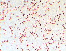


Photo Gallery Of Pathogenic Bacterial


Laboratory Science Review Microbiology Medical Laboratory Science Medical Laboratory


Escherichia Coli E Coli An Overview Microbe Notes


Module 7 8 10 Gram Stain Acid Fast Endospore Growth Characteristics Flashcards Quizlet


Solved For Each Image Describe The Bacteria S Color The Chegg Com


Staining Microscopic Specimens Microbiology


Recombinant Production Of The Therapeutic Peptide Lunasin Microbial Cell Factories Full Text


Gram Staining Principle Procedure And Results Msc Sarah Ahmed Ppt Download


Laboratory Perspective Of Gram Staining And Its Significance In Investigations Of Infectious Diseases Thairu Y Nasir Ia Usman Y Sub Saharan Afr J Med


Escherichia Coli Wikipedia


E Coli In Gram Stain Introduction Pathogenic Strains And Lab Diagnosis


Morphology Culture Characteristics Of Escherichia Coli E Coli


Gram Stain Of E Coli Bacterium A Gram Stain Of Shows Gramnegative Download Scientific Diagram


Bacterial Staining


Escherichia Coli Morphology Mixed Rods Gram Stain Gram Negative Bacilli Growth Characteristics Dubai Khalifa


Gram Stain


Gram Stained Images Of E Coli Lower Panels And B Subtilis Upper Download Scientific Diagram


コメント
コメントを投稿